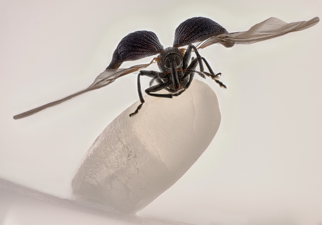
Balancing Weevil Wins 2025 Nikon Small World Photo Contest
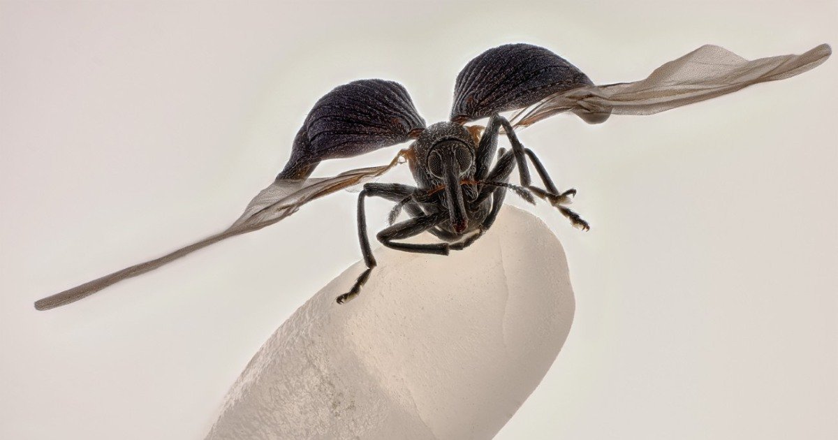
Zhang Yu
Kunming, Yunnan, China, 1st place, Rice weevil (Sitophilus oryzae) on a grain of rice, image stack, 5X (objective lens zoom)
It may look like an alien perched on a crystal, but the winning photo is by Zhang You from Nikon’s small world Competition is not as evil as it seems. In fact, an insect lover snapped a stunning photo of a rice weevil sitting on a grain of rice. It’s stunning images like these that make Nikon Small World so unique in the world of photography competitions.
Launched in 1974, the competition highlights the world of scientific microscopy and digital photography. By introducing these images to a wider audience, they help the public understand how artistic microscopic photography can be. For example, Zhang spent years perfecting his craft by examining insect behavior, and his winning photo shows the benefits he gained from this practice.
“It is useful to delve into entomology: understanding insect behaviour, mastering lighting,” he says. “The outstanding work blends artistry with scientific rigor, capturing the essence, energy and spirit of these creatures.”
Interestingly, this was Zhang’s first time entering the competition, and not only did he win, but he also secured another spot in the top 20 list.
“His achievement highlights the Nikon Small World ethos: inspiring wonder, making scientific understanding accessible to all, and celebrating the ingenuity of the microscopic field,” said Eric Flem, senior director of communications and CRM at Nikon Instruments.
Other key images include everything from pollen grains trapped in a spider’s web to the muscles of heart cells, demonstrating the diversity of what can be imaged under a microscope.
The announcement of these winners follows Nikon is a small world in motion A competition that awards excellence in moving images captured with a microscope. Combined, they both offer a revealing look at the unseen world that surrounds us.
Here are the 12 best photos from the 2025 Nikon Small World Photo Contest.
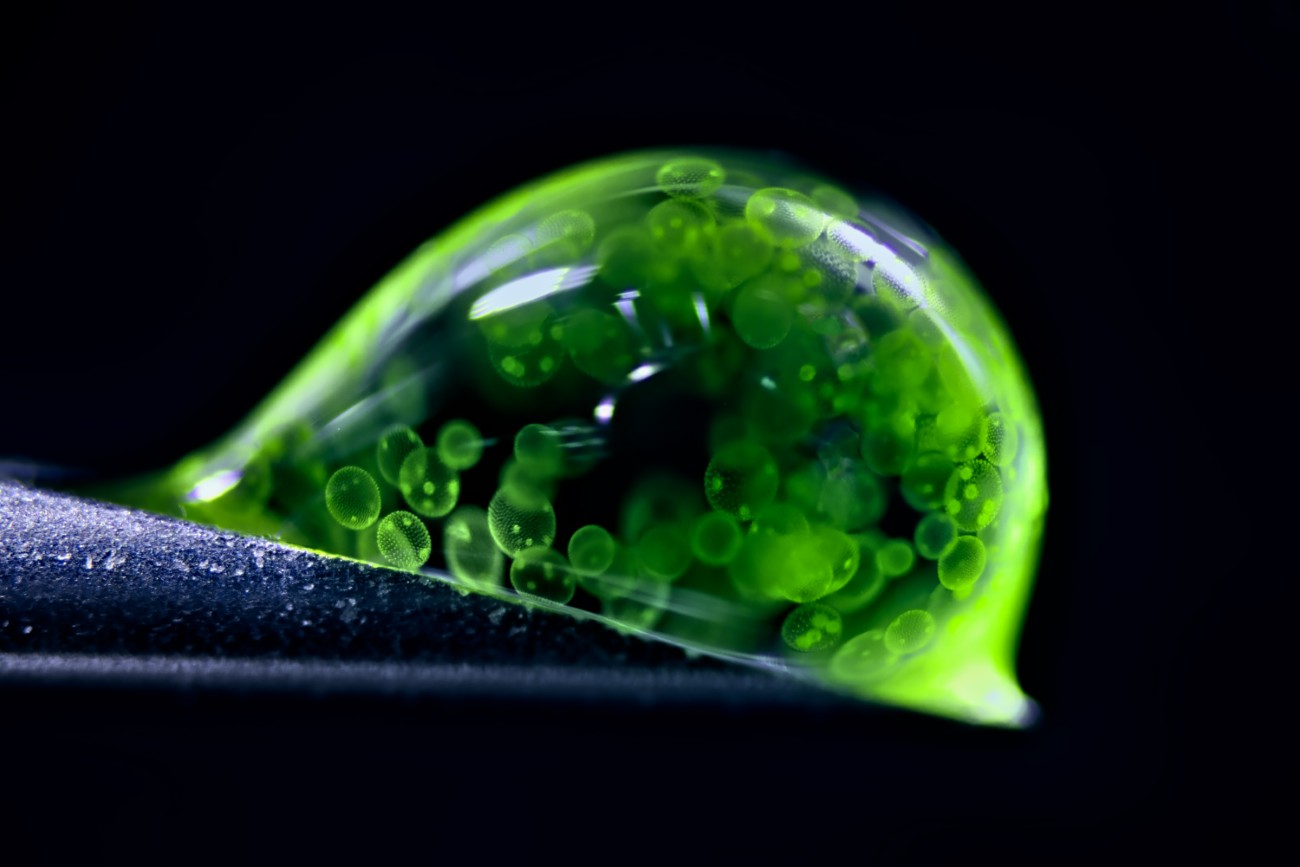
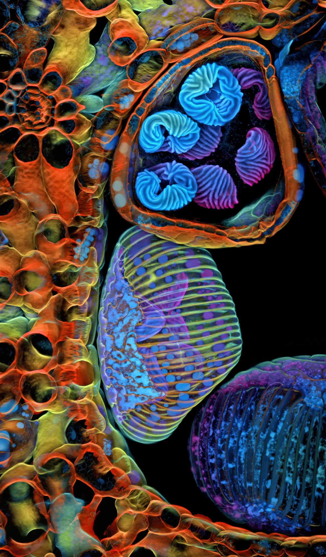
Dr. Jan Rosenbaum
Rostock, Mecklenburg-Vorpommern, Germany, 2nd place, Colonial algae (Folvox) in a drop of water, reflected light, 5X (objective lens magnification)

John Oliver is stupid
Medienbunker Production, Bendorf, Rhineland-Palatinate, Germany, 3rd place, pollen in a garden spider web, image stack, 20X (objective lens magnification)
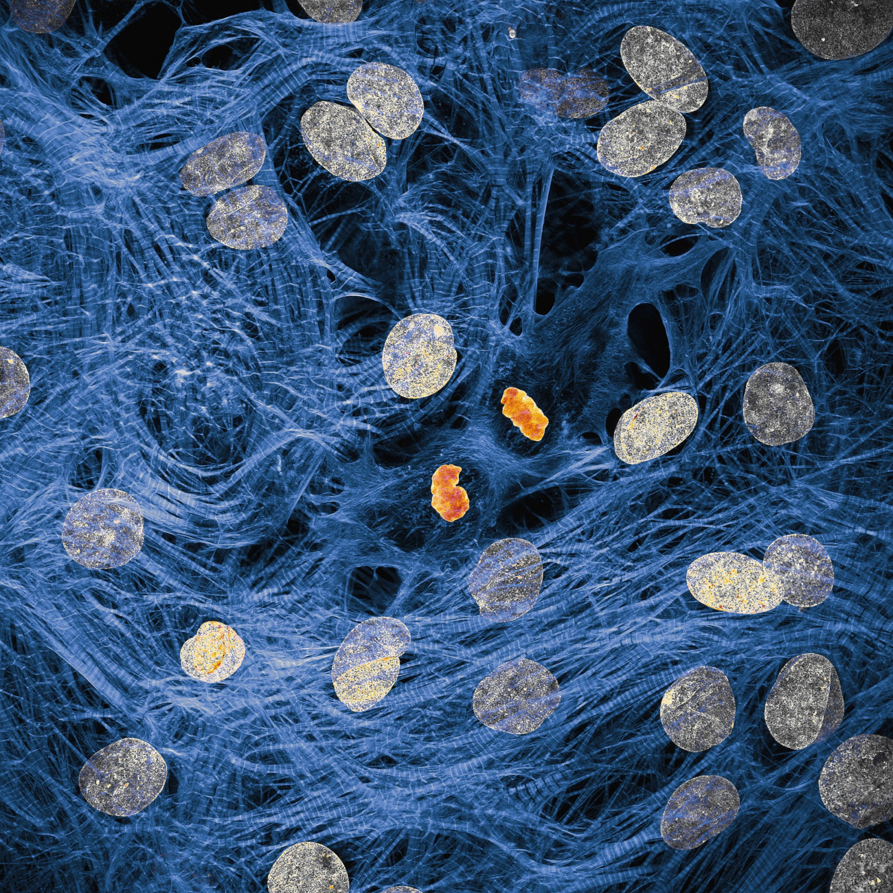
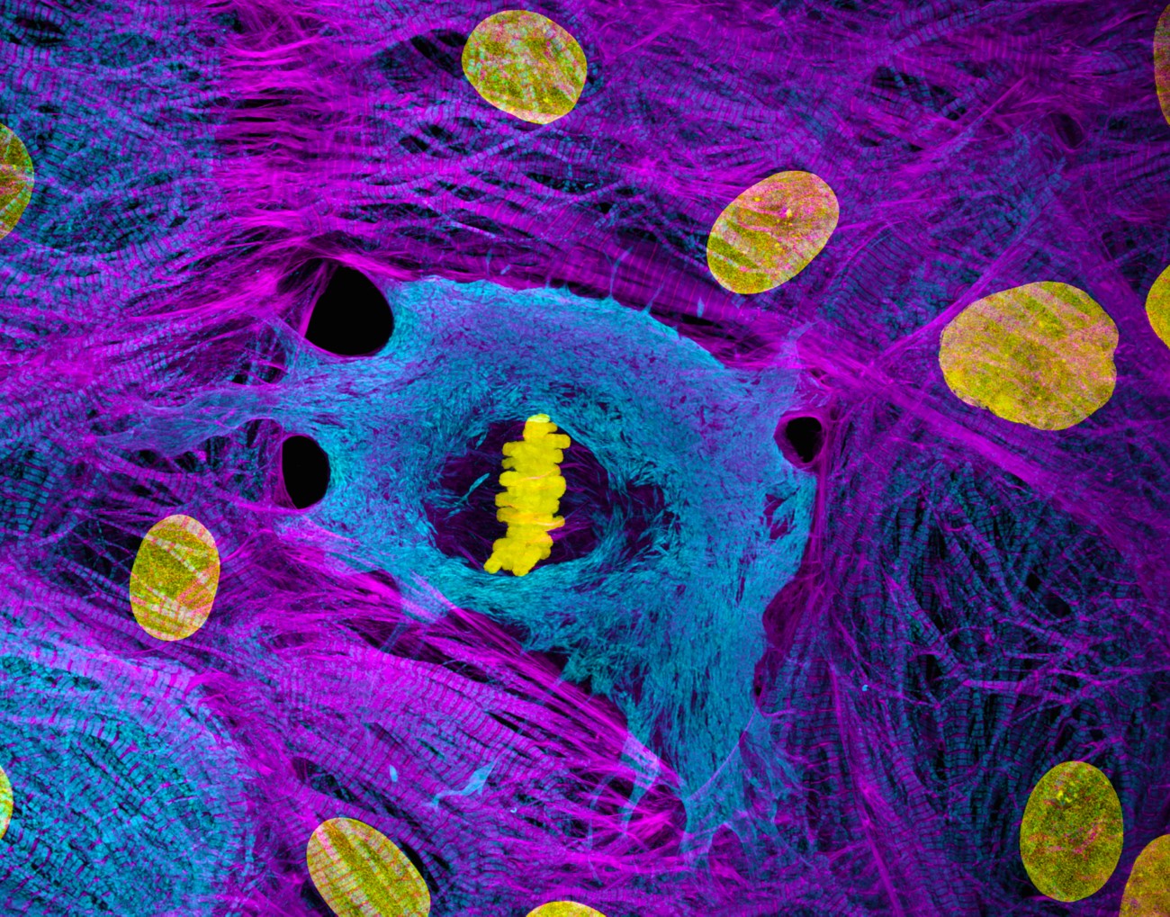
Dr. James Hayes, Vanderbilt University, Department of Cell and Developmental Biology, Nashville, TN, USA, IV, Cardiomyocytes with post-cell division chromosomal condensation, confocal, 100X (objective lens magnification)
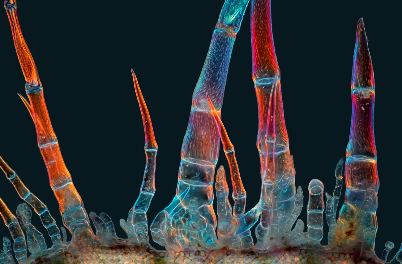
Dr. Igor Siwanovich, Howard Hughes Medical Institute (HHMI), Janelia Research Park, Ashburn, VA, USA, 5th Place, Spores (blue/purple structures) of a small tropical fern (Ceratopteris richardii), confocal, 25X (objective lens magnification)
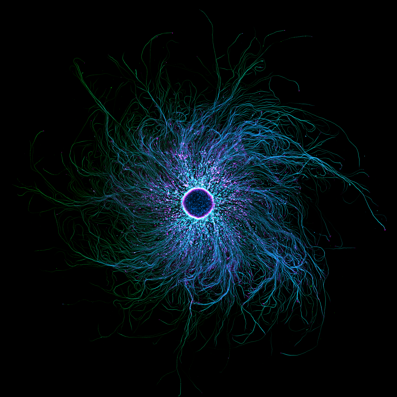
Stella Whitaker, National Institutes of Health, National Institute of Neurological Disorders and Stroke
Bethesda, Maryland, USA, 7th, iPSC-derived sensory neurons labeled to show tubulin and actin, confocal, fluorescence, image stacking, 10X (objective lens magnification)
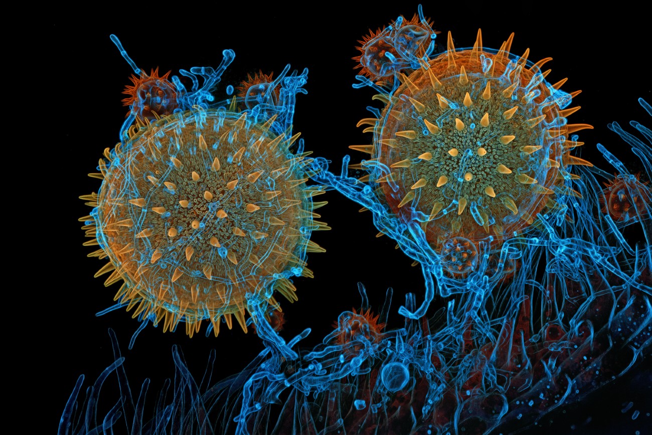
Dr. Igor Siwanovich, Howard Hughes Medical Institute (HHMI), Janelia Research Campus
Ashburn, Virginia, USA, 8th, Mallow pollen sprouting on stigma while parasitized by a filamentous fungus, confocal, 40X (objective lens magnification)
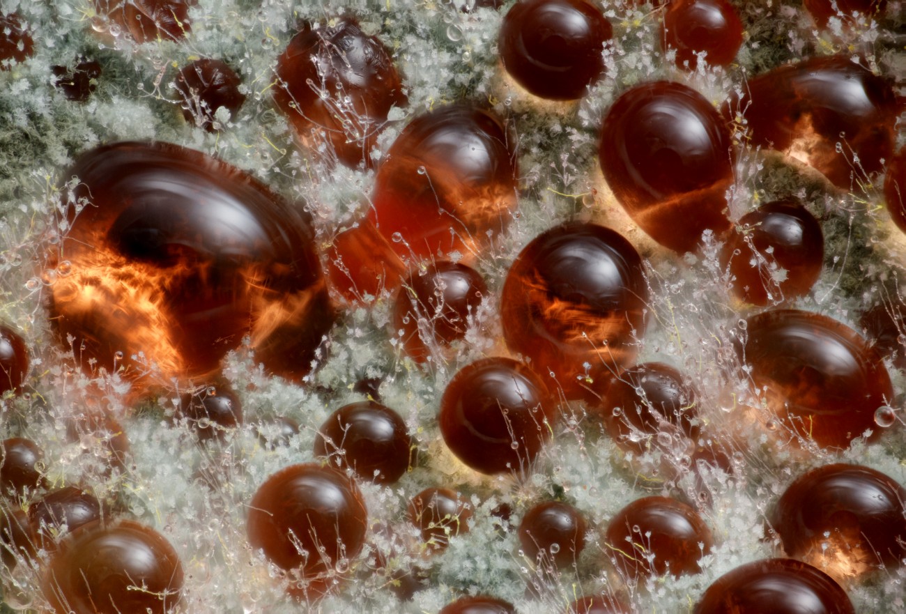
Wim van Egmond, Micropolitan Museum, Berkel en Rodenris, Zuid Holland, Netherlands, 9th, Mushroom (Talaromycespurpureogenus) known for its diffuse red pigment, image stack, 10X (objective lens magnification)
Dr. Dylan Burnett and Dr. James Hayes, Vanderbilt University School of Medicine
Department of Cell and Developmental Biology, Nashville, TN, USA, 10th, Cardiomyocytes (iPSC-derived) showing condensed chromosomes at metaphase, structured illumination, microscopy (SIM), 60X (objective lens magnification)
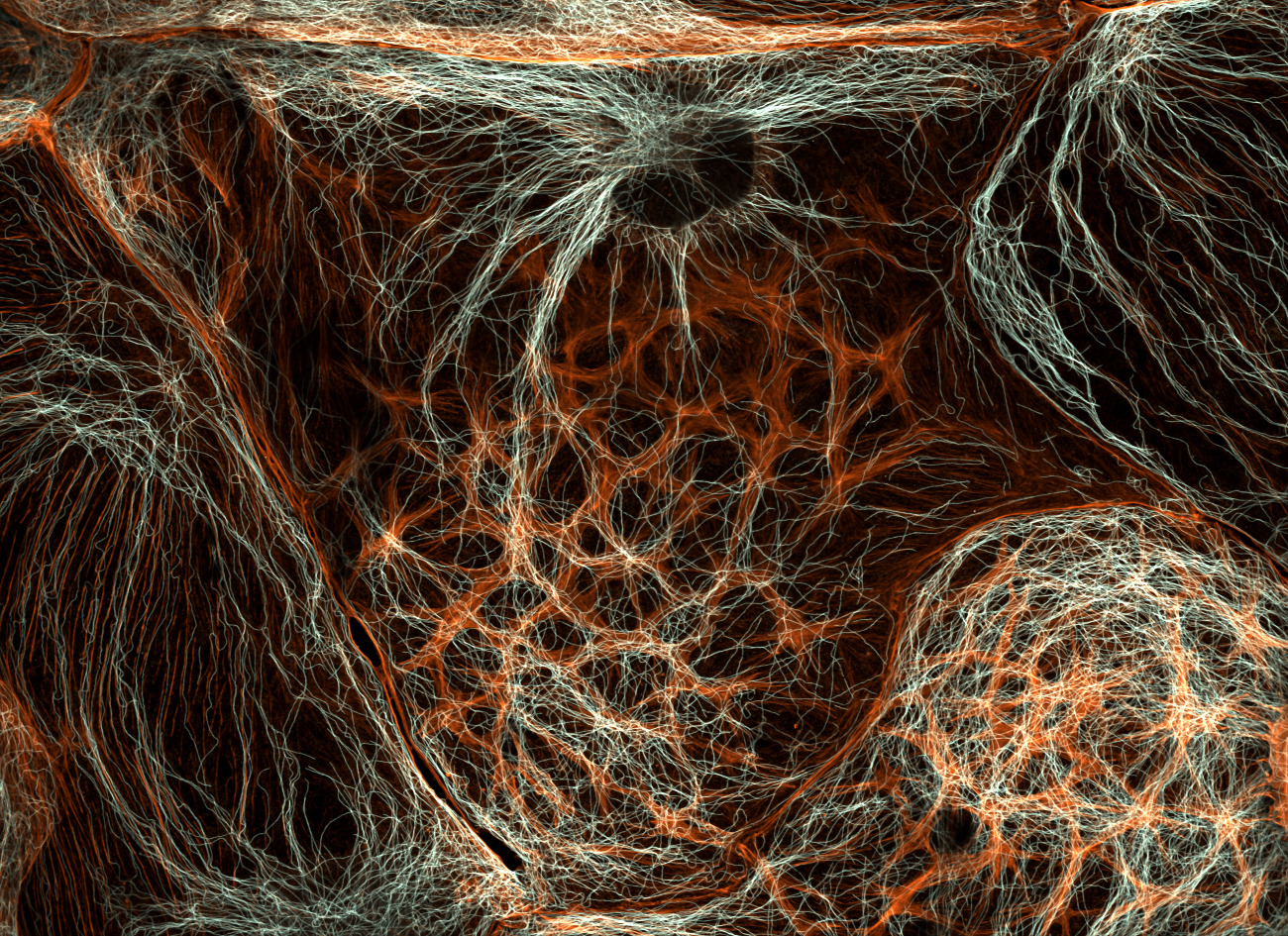
Dr. Francisco Lazaro-Dieguez, Albert Einstein College of Medicine, Bronx, NY, USA, VI, rat liver cells, confocal, 63X (objective lens magnification)
Marek Miś, Marek Miś Photography, Suwalki, Podlaskie, Poland, 11th place, trichromatic sunflower (hair-like plant growth), polarized light, 10X (objective lens magnification)
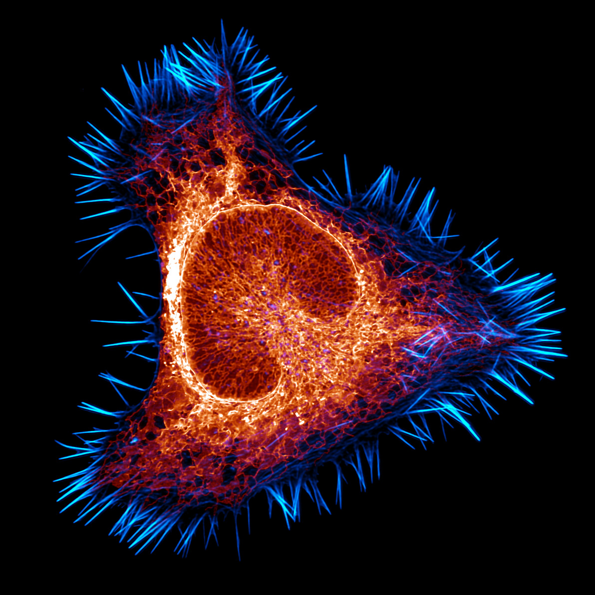
Haley Lindamood and Eric Vitriol, Augusta University, Department of Neuroscience and Regenerative Medicine
Augusta, Georgia, USA, 12th, Actin cytoskeleton (cyan) and endoplasmic reticulum (red) of a mouse brain cancer cell, confocal, deconvolution, 100X (objective lens magnification)
Nikon small world: Website | Facebook | Instagram
My Modern Met has granted permission to display images by Nikon Small World.
Related articles:
Wildlife photography contest honors nature’s resilience
A rare brown hyena photo wins the 2025 Wildlife Photographer of the Year competition
Colorful ‘sea beetles’ win the 2025 Ocean Photographer of the Year competition
A bird flying under a total solar eclipse wins the 2025 Bird Photographer of the Year award
Share this content:


Post Comment